Lung Parenchyma Location : Lung Anatomy Physiopedia / The most prominent structure in this region is the alveolus (figure 1).
byAdmin-
0
Lung Parenchyma Location : Lung Anatomy Physiopedia / The most prominent structure in this region is the alveolus (figure 1).. But in interstitial lung disease, the repair process goes awry and the tissue around the air sacs (alveoli) becomes scarred and thickened. The closest locations of metastases of the primary lesions are in the peritoneum and bowels 7, 8. Arises most commonly from pleura, with uncommon inward growth into lung parenchyma extremely rare to arise entirely within lung parenchyma generally peripheral in location pathophysiology. Consolidation occurs when the normally air filled lung parenchyma becomes engorged with fluid or tissue, most commonly in the setting of pneumonia. Rarely, pulmonary nodules are a sign of lung cancer.
Parenchyma the parenchyma of the pineal body is comprised of lobules with a definite cytoarchitectural pattern and surrounded by connective tissue septa (fig. The most prominent structure in this region is the alveolus (figure 1). The main difficulty comes in describing abnormalities of the lung parenchyma. The other main type of. The left side of the screen is lung parenchyma (remember that left is cephalad and right is caudad).
Pneumonia Acute Inflammation Of The Lung Parenchyma The from s2.studylib.net What some may call 'shadowing,' others may call 'opacification,' 'whiteness,' or 'increased density.'. The percentage is higher on computed tomography which can detect disease when the radiograph is normal. The lobules contain 3 varieties of elements: The most prominent structure in this region is the alveolus (figure 1). Lung parenchyma is the substance of the lung that is involved with gas exchange and includes the pulmonary alveoli and respiratory bronchioles, though some authors include only the alveoli. Interstitial lung disease seems to occur when an injury to your lungs triggers an abnormal healing response. In general, squamous carcinomas are encountered. The most accurate way to determine if a lung disease affects this part of the lung is with a surgical biopsy.
Parenchyma the parenchyma of the pineal body is comprised of lobules with a definite cytoarchitectural pattern and surrounded by connective tissue septa (fig.
Lung parenchyma is the substance of the lung that is involved with gas exchange and includes the pulmonary alveoli and respiratory bronchioles, though some authors include only the alveoli. Bird fancier's lung (avian proteins) is most common in children! An umbrella term that encompasses a large number of disorders that are characterised by diffuse cellular infiltrates in a periacinar location. Patients usually respond to removal of antigen. The lung parenchyma is further subdivided. The closest locations of metastases of the primary lesions are in the peritoneum and bowels 7, 8. In general, squamous carcinomas are encountered. If a large enough segment of parenchyma is involved, it can alter the transmission of air and sound. Most lung nodules are benign (not cancerous). Parenchyma in the lungs essentially includes all systems and tissues pertinent to the lung's healthy functioning. Infected lung parenchyma can affect a person's breathing. Pneumonia is an infection of the lung parenchyma caused by a wide variety of organisms in pediatric patients. The lung parenchyma is that portion of the lungs involved in gas exchange.
The liver parenchyma is the functional tissue of the organ made up of around 80% of the liver volume as hepatocytes. Respiratory area of the lungs (lung parenchyma) the structures in the lungs directly responsible for the function of respiration collectively form the lung parenchyma. Roughly half of people who smoke over the age of 50 will have nodules on a ct scan of their chest. The percentage is higher on computed tomography which can detect disease when the radiograph is normal. What some may call 'shadowing,' others may call 'opacification,' 'whiteness,' or 'increased density.'.
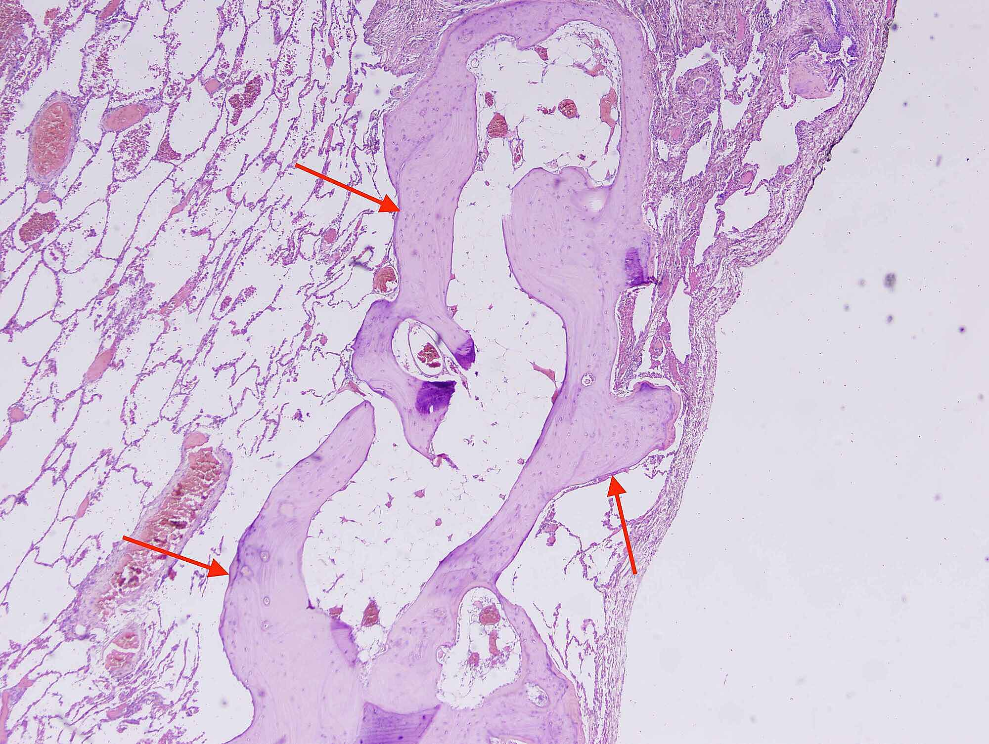
Cureus First Case Of Dendriform Pulmonary Ossification In Bahrain from assets.cureus.com Patients usually respond to removal of antigen. A lung (pulmonary) nodule is an abnormal growth that forms in a lung. The percentage is higher on computed tomography which can detect disease when the radiograph is normal. Lung parenchyma is the substance of the lung that is involved with gas exchange and includes the pulmonary alveoli and respiratory bronchioles, though some authors include only the alveoli. Brody, md cincinnati children's hospital. Diffuse parenchymal lung diseases are disorders that affect the interstitial of the lungthe area around the lung's air sacs. Pneumatosis is also a frequent result of surgery. The closest locations of metastases of the primary lesions are in the peritoneum and bowels 7, 8.
This effectively rules out pleural effusion at this location.
Or a lump in a surgical patient. Interstitial lung disease seems to occur when an injury to your lungs triggers an abnormal healing response. No cartilage or rib excision had taken place in the previous operation. But in interstitial lung disease, the repair process goes awry and the tissue around the air sacs (alveoli) becomes scarred and thickened. Lung nodules are being recognized more frequently with the wider application of ct screening for lung cancer. Lung parenchyma, however, more extensively involves the bronchioles or lung airways, as well as key blood vessels located inside of the lungs. Parenchyma in the lungs essentially includes all systems and tissues pertinent to the lung's healthy functioning. We report the ct findings of parenchymal and pleural diseases in a group of patients with a history of asbestos exposure, excluding lung cancer (which is not typical in this subjects) and asbestosis (which is a parenchymal fibrosis). The closest locations of metastases of the primary lesions are in the peritoneum and bowels 7, 8. Each alveolus in the lung parenchyma opens directly into an alveolar duct or occasionally, in a limited number of species, into a respiratory bronchiole. Lung parenchyma is the substance of the lung that is involved with gas exchange and includes the pulmonary alveoli and respiratory bronchioles, though some authors include only the alveoli. Attention should be given to factors such as location, size, shape and density of an abnormality. Lung anatomy includes the lung parenchyma, which carries part of the conduction system but is mainly involved in the gas exchange at the alveolar level.
Parenchyma the parenchyma of the pineal body is comprised of lobules with a definite cytoarchitectural pattern and surrounded by connective tissue septa (fig. Lung parenchyma, however, more extensively involves the bronchioles or lung airways, as well as key blood vessels located inside of the lungs. Diffuse parenchymal lung diseases are disorders that affect the interstitial of the lungthe area around the lung's air sacs. Ordinarily, your body generates just the right amount of tissue to repair damage. What some may call 'shadowing,' others may call 'opacification,' 'whiteness,' or 'increased density.'.
Multiple Lung Parenchymal Abnormalities Don T Panic Let S Be Pragmatic The 6 Question Rule A Checklist Strategy Sciencedirect from ars.els-cdn.com Respiratory area of the lungs (lung parenchyma) the structures in the lungs directly responsible for the function of respiration collectively form the lung parenchyma. Consolidation occurs when the normally air filled lung parenchyma becomes engorged with fluid or tissue, most commonly in the setting of pneumonia. Each alveolus in the lung parenchyma opens directly into an alveolar duct or occasionally, in a limited number of species, into a respiratory bronchiole. Diffuse parenchymal lung diseases are disorders that affect the interstitial of the lungthe area around the lung's air sacs. Interstitial lung disease seems to occur when an injury to your lungs triggers an abnormal healing response. It includes the bronchial tubes, blood vessels, alveoli, and alveolar ducts 31, 32. Lung parenchyma, however, more extensively involves the bronchioles or lung airways, as well as key blood vessels located inside of the lungs. The most accurate way to determine if a lung disease affects this part of the lung is with a surgical biopsy.
Ordinarily, your body generates just the right amount of tissue to repair damage.
As the patient inspires, a curtain of grey air artifact sweeps from left (superior) to right (inferior) obliterating the view of the diaphragm and liver. Pneumonia is an infection of the lung parenchyma caused by a wide variety of organisms in pediatric patients. Parenchyma in the lungs essentially includes all systems and tissues pertinent to the lung's healthy functioning. Each alveolus in the lung parenchyma opens directly into an alveolar duct or occasionally, in a limited number of species, into a respiratory bronchiole. Or a lump in a surgical patient. Arises most commonly from pleura, with uncommon inward growth into lung parenchyma extremely rare to arise entirely within lung parenchyma generally peripheral in location pathophysiology. Lung parenchyma is the substance of the lung that is involved with gas exchange and includes the pulmonary alveoli and respiratory bronchioles, though some authors include only the alveoli. The percentage is higher on computed tomography which can detect disease when the radiograph is normal. If a large enough segment of parenchyma is involved, it can alter the transmission of air and sound. The most prominent structure in this region is the alveolus (figure 1). Pneumoperitoneum (or peritoneal emphysema) is air or gas in the abdominal cavity, and is most commonly caused by a perforated abdominal organ. Attention should be given to factors such as location, size, shape and density of an abnormality. If lung tissue is obtained, however, there is histologic disease in almost all patients, including those who have
An umbrella term that encompasses a large number of disorders that are characterised by diffuse cellular infiltrates in a periacinar location lung parenchyma. Pneumoperitoneum (or peritoneal emphysema) is air or gas in the abdominal cavity, and is most commonly caused by a perforated abdominal organ.